Optimization of MRI Protocol
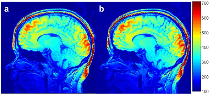
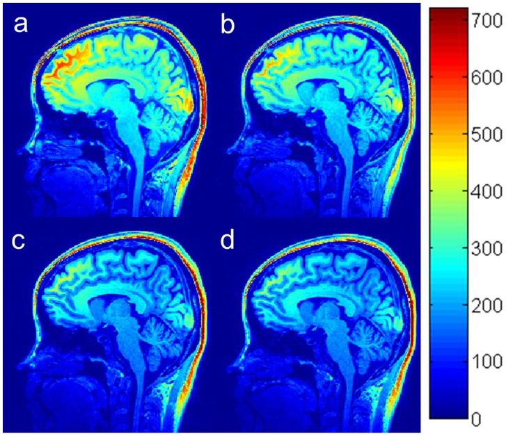
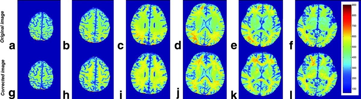
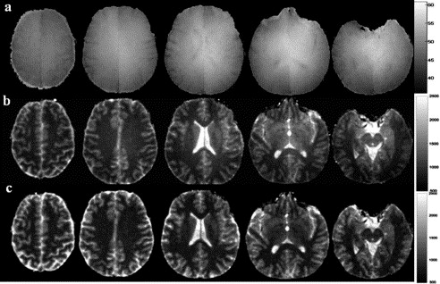
Description: A standard protocol for brain imaging may include T1-weighted, T2-weighted, Fluid-attenuated inversion recovery (FLAIR), diffusion weighted, susceptibility weighted or T2*-weighted, and functional MRI imaging. Three dimensional (3D) T1-weighted imaging is one of the most popular sequences for structural brain imaging in clinical and research settings. The existing imaging parameters of 3D T1-weighted imaging is general. It is sub-optimal for specific scanner and sub-disease group. I proposed a method, with a team of collaborators, to optimize the imaging parameters for a given scanner for reaching fullest potential. That is, the imaging parameters were personalized for a given k-space trajectory and contrast agent. These studies greatly improved and increased signal-to-noise efficiency and contrast-to-noise efficiency up to 50% with or without the administration of contrast agent. The outcome holds potential to personalize and optimize the brain imaging protocol. It allows us to apply more valuable MRI scan time for clinical and research applications.
Reference:
- Wang J, He L, Zheng H, Lu ZL. Optimizing the magnetization-prepared rapid gradient-echo (MP-RAGE) sequence. PLoS One. 2014;9(5):e96899. PMID: 24879508 Pubmed Journal
- Wang J, He L, Zheng H, Lu ZL. Improving structural brain images acquired with the 3D FLASH sequence. Magn Reson Imaging. 2017;38:224-232. PMID: 28109888 Pubmed Journal
- Wang J, Lu ZL (2016). Methods and apparatus for optimization of MRI protocol. (Issued, US patent 9339239 B2). Pubmed Journal
- Wang J, Parikh N, Lu ZL, He L (2018). Methods and apparatus for optimization of MRI protocol. (US pending, US 2018/0045799 A1). Pubmed Journal