Diffuse White Matter Abnormalities Segmentation
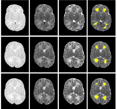
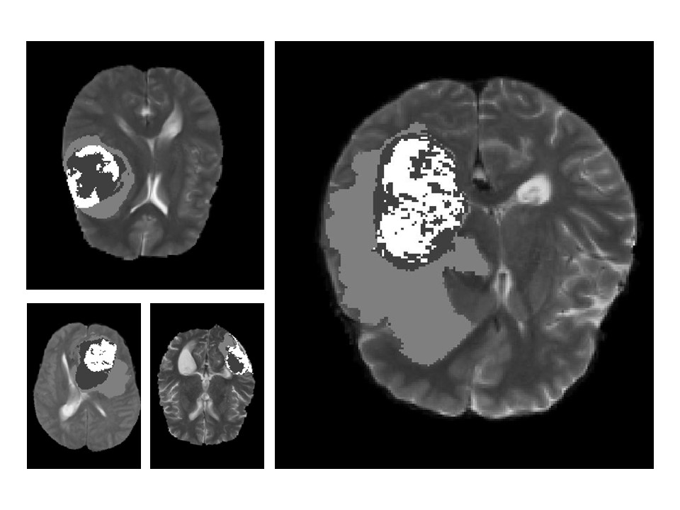
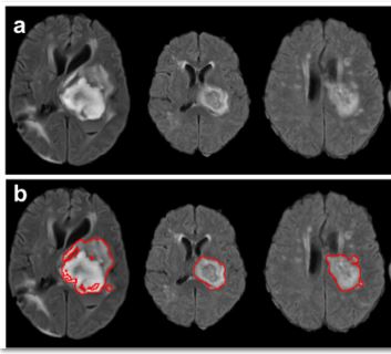
Introduction: Diffuse white matter abnormality (DWMA) is observed in 50-80% of very preterm infants at term-equivalent age. It is characterized by either 1) diffusely higher signal intensity in periventricular and subcortical white matter than in normal unmyelinated white matter on T2-weighted MRI images (also known as diffuse excessive high signal intensity; or 2) lower signal intensity than unmyelinated white matter on T1-weighted and fluid-attenuated inversion recovery (FLAIR) sequences.
Despite the high prevalence of DWMA, the pathological significance of DWMA continues to be debated. Several neurodevelopmental follow-up studies have revealed that DWMA is predictive of lower developmental scores. Nevertheless, others have not observed significant links between DWMA and neurodevelopmental impairments. Much of this debate has been fueled by the nearly universal use of qualitative reporting of DWMA that is subjective and unreliable, likely resulting in measurement error and lack of association with neurodevelopmental impairments in some studies. Thus, accurate and automatic detection of DWMA is of crucial importance for understanding DWMAs biologic nature and potentially risk stratifying high-risk preterm infants that may benefit from early intervention therapies.
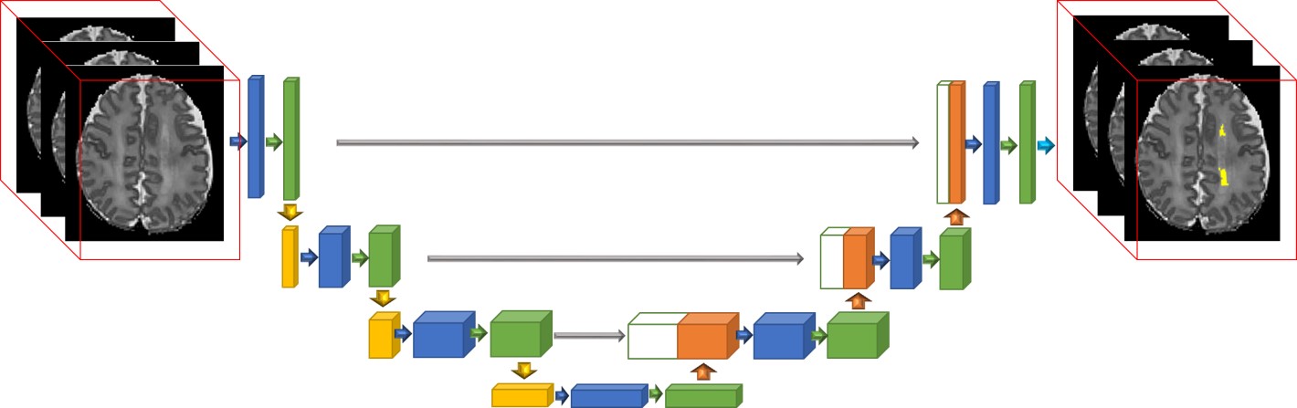
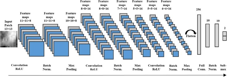
Utilizing the local neighboring spatial information of individual voxels, we proposed a sequence of automated and objective DWMA segmentation approaches, including the advanced deep convolutional neural networks and 3D Residual U-Net models, to identify DWMA regions on T2-weighted MRI images for very preterm infants. (He and Parikh 2013, He and Parikh 2013, Li, Parikh et al. 2019) With our DWMA segmentation methods, we found that objectively diagnosed DWMA is an independent predictor of cognitive, language, and motor development in very preterm infants. (Parikh, Harpster et al. 2020, Parikh, He et al. 2020)
Reference:
Parikh NA, He L, Priyanka Illapani VS, Altaye M, Folger AT, & Yeates KO. (2020). Objectively Diagnosed Diffuse White Matter Abnormality at Term Is an Independent Predictor of Cognitive and Language Outcomes in Infants Born Very Preterm. J Pediatr, 220, 56-63. PMID: 32147220. Pubmed Journal
Parikh N.A., Harpster K., He L., Priyanka Illapani VS., Khalid F.C., Klebanoff M.A., O'Shea T.M., Altaye M. (2020) Novel diffuse white matter abnormality biomarker at term-equivalent age enhances prediction of long-term motor development in very preterm children. Sci Rep 10, 15920 (2020). https://doi.org/10.1038/s41598-020-72632-0 Pubmed Journal
Li H., Parikh N.A., Wang J., Merhar S., Chen M., Parikh M., Holland S., He L., (2019). Objective and automated detection of diffuse white matter abnormality in preterm infants using deep convolutional neural networks. Frontiers in Neuroscience, in press. PMC Journal
He L, & Parikh NA. (2013). Automated detection of white matter signal abnormality using T2 relaxometry: application to brain segmentation on term MRI in very preterm infants. Neuroimage, 64, 328-340. PMID: 22974556; PMCID: PMC3544934. PMC Pubmed Journal